
|
Author(s)
David Miller
Keith Harding
|
Contents
|
|
Published:
Dec 2003
Last updated: November 2009 Revision: 1.2 |
Keywords: pilonidal; abscess; surgery; sinus; infection.
Pilonidal disease is a common complaint affecting mainly men from puberty to their early thirties.
Acute abscesses need to be drained and followed up with meticulous attention to hair and debris removal and wound packing.
Wound infection should be treated with broad spectrum antibiotics, such as metronidazole and erythromycin, for at least two weeks depending upon response.
Chronic pilonidal sinuses are best treated with wide excision and secondary healing.
Long-term recurrence is reduced by good hygiene and regular depilation.
A pilonidal sinus (PNS) occurs in the cleavage between the buttocks (natal cleft) and can cause discomfort, embarrassment and absence from work for thousands of young people (mostly men) annually. It is a common problem in primary care due to recurrence following surgery and the need for frequent and time-consuming wound care. This article covers the pathology, clinical presentations and appropriate management of pilonidal sinus disease.
Pilonidal disease was first described by Hodges in 1880 [1] and is diagnosed by the finding of a characteristic epithelial track (the sinus) situated in the skin of the natal cleft, a short distance behind the anus and generally containing hair, hence the name pilonidal taken from the Latin, meaning literally 'nest of hairs'. During the Second World War the condition was common in jeep drivers, which led to it being known as 'jeep disease'. A similar condition arises in the clefts between the fingers of barbers or hairdressers caused by customers' hair entering moist, damaged skin.
The onset of PNS is rare both before puberty and after the age of 40. Males are affected more frequently than females, probably due to their more hirsute nature [2]. In a population study of 50,000 students the incidence in males was 1.1%, ten times more than in females [3], although many of these were asymptomatic. Figures from the Office of Population Censuses and Surveys (1985) record 7,000 patients as having had inpatient treatment for PNS in England. Proportionately females make up a quarter of those undergoing hospital treatments [4], possibly reflecting under-reporting in the male population. The condition is more common in Caucasians than Asians or Africans due to differing hair characteristics and growth patterns [5]. In a study of risk factors the following associations were found [2]:
sedentary occupation 44%
positive family history 38%
obesity 50%
local irritation or trauma prior to onset of symptoms 34%.
The origin of pilonidal disease is not fully understood, but the majority of opinion favours the acquired theory, which may be summed up as:
Hormones
Hair
Friction
Infection
Sex hormones first produced at puberty are known to affect the pilosebaceous glands, which coincides with the earliest onset of pilonidal disease [6]. Pilonidal disease is associated with visible pits in the midline of the natal cleft, which have the microscopic appearance of enlarged hair follicles[7]. The enlargement is thought to occur due to stretching of the follicular openings caused by the weight of the buttocks being pulled by gravity[7]. The magnitude of this force is enhanced by its application over a small area (approximately 1mm²), where the natal cleft curves over the sharp angle at the sacro-coccygeal joint. This force may be amplified by activities such as bouncing while upright, slumping while seated or bouncing on a hard seat as in 'jeep disease'. If the force applied reaches a critical level then the base of the follicle ruptures as this is the weakest part at which the skin is tethered to the subcutaneous tissue. The same forces, together with friction between the buttocks, are also responsible for sucking keratin and hair into the distended follicles, which leads to infection of the follicle and abscess formation.
It had long been believed that hair follicles alone were the source of pilonidal disease[7]. However, more recent work on specimens following wide excision has shown that the pits penetrating into the dermis were distended with keratin and debris, but not all arose in hair follicles[8].
Furthermore, hair entry to the follicles by a variety of means was originally thought to be the primary event in the development of PNS. However, there is evidence to suggest that the enlargement of the follicles precedes hair gaining access and at operation hair is found in only half of cases[7]. Hair acts as a foreign body causing an inflammatory reaction and can lead to prolonged inflammation and the development of chronic pilonidal disease. As such, the hairs that gave pilonidal disease its name may not be the primary insult but nonetheless are an important secondary event. The source of hair can be either the natal cleft itself in hirsute individuals, or hair from the head or back that falls down inside clothes into the natal cleft. Distension of the obstructed follicle leads to oedema and inflammation resulting in occlusion of the mouth of the pit, with subsequent infection and abscess formation in the subcutaneous fatty tissue (the patient may present at this stage). Over time a chronic abscess develops and it is the formation of the track draining this abscess cavity that is known as a sinus. The combination of a deep abscess cavity with surrounding moist conditions and abundant bacteria, hair, debris and friction cause recurrent infection, associated with chronic pain and discharge.
Malignant change is a relatively rare complication of pilonidal disease[8], but evidence of at least 50 such cases is available in published medical literature. The most common scenario is of squamous cell carcinoma arising after decades of antecedent pilonidal disease. Its pathophysiology is uncertain, but the ongoing processes of tissue damage and repair in the presence of chronic inflammation may be implicated[9]. Malignancy arising in a chronic wound seems to have a worse prognosis than cutaneous malignancies arising de novo on the skin, hence early detection is imperative[9]. Although malignancy is a rare complication, it is a significant risk to young men and a more sinister reason why, besides discomfort and inconvenience, pilonidal disease should be taken seriously and sufferers encouraged to seek medical help.
Anaerobic bacteria (particularly Bacteroides and Enterococci) predominate over aerobic bacteria (Staphylococci and Haemolytic Streptococci) in the development of infection of the follicles and abscess formation. Anaerobes are predominantly responsible for reinfection and subsequent wound breakdown following surgery[10]. However a more recent prospective study of 49 infected postoperative wounds isolated aerobic bacteria in 43% of cases[11]. Treatment of pilonidal infection should be with broad spectrum antibiotics.
A PNS may be asymptomatic for some time prior to presentation. The majority of patients only present with the onset of symptoms, usually pain and discharge. Occasionally a painless lump or swelling may be discovered by the patient while washing, or the characteristic midline pits may be found during a routine physical examination. Symptomatic disease usually presents[12] as an acute pilonidal abscess, a chronic pilonidal abscess or complex/recurrent pilonidal disease.
The patient notices increasing discomfort and swelling over a number of days and the pain may be severe by the time of presentation. On examination there is a localised fluctuant swelling in the midline of the natal cleft with overlying cellulitis. The area is exquisitely painful to touch and often simply the act of separating the buttocks to examine the area is intolerable for the patient.
It is common for patients to present with chronic pain and discharge, often with a history of up to two years[2]. On examination a single, or occasionally, multiple sinuses may be seen. Tufts of hair or other debris, such as clothing fibres, are often visible arising from the sinus. Localised oedema, swelling and inflammation may be present masking the underlying sinus.
This is due to reinfection in neighbouring hair follicles or chronic infection from entry of hair or debris into a postoperative wound.
This will depend upon the nature of presentation and the attitude and wishes of the individual concerned. Regardless of the treatment selected, the aims remain similar:
Treatment must be acceptable to the patient in terms of discomfort, impact upon body image and self-esteem
Rates of complication and recurrence should be minimal
To enable the patient to resume work and normal social activities as early as possible.
For those patients who are truly asymptomatic, meticulous depilation and local hygiene are advised. It is not known what proportion of those who are asymptomatic go on to develop symptomatic disease. However, the risks and discomfort of elective surgery for most of these patients would be unacceptable and probably outweigh the potential benefit of avoidance of future abscess formation. Hence surgical intervention in this group is not advised.
For the acute abscess that presents early, with pain that is tolerable and no evidence of cellulitis, broad spectrum antibiotics and depilation alone may be sufficient to resolve the immediate problem. If symptoms resolve, follow-up examination for the presence of pits or sinus tracks is recommended along with long-term depilation and attention to hygiene. The majority of acute cases do, however, require urgent operative intervention. When pain is severe or cellulitis is present, attempts at conservative treatment are likely to be futile and will only prolong the patient's discomfort.
Chronic disease can be successfully treated by shaving and meticulous hygiene, but recurrence rates are unknown[13].
This is a closed technique under local anaesthetic whereby injection of phenol into a sinus causes sclerosis and gradual closure[14]. The procedure is time consuming, needs frequent repetition, has a high recurrence rate and has been largely replaced by operative techniques.
The number and variety of published techniques are testament to the complexity of treating PNS and the fact that no single procedure is superior in all respects. It is universally agreed that the most effective emergency management of a pilonidal abscess is simple incision and drainage. However, surgical management of chronic and recurrent disease is more controversial. Numerous studies have been put forward advocating one treatment over another, but many of these studies are weighed down by lack of control groups or short follow-up. The majority of procedures can be classified in one of the four categories below:
Incision and drainage
Excision and healing by secondary intention
Excision and primary closure
Excision with reconstructive flap techniques.
Incision and drainage: This is a simple procedure that involves making an elliptical incision in the abscess just off the midline. The mouth of the wound should be of sufficient width to allow packing of the entire wound cavity. Curettage to remove dead or infected tissue in the wound improves the rate of healing, with 90% completely healed at one month, compared to just 58% healed at 10 weeks in the absence of curettage[5].
Following a single incision and drainage procedure, 40-60% will go on to develop a PNS requiring further surgery. Pits or sinuses can be excised as part of an incision and drainage procedure, but these can be obscured by oedema and are often overlooked at the initial assessment[7]. The recurrence rate can be reduced to about 15% if a second procedure to excise pits and sinuses is performed after five to seven days[5].
Healing by secondary intention has the advantage of allowing free drainage of infected material and debris. However the patient will require regular wound care and the discomfort of packing until the wound has closed. In a retrospective study mean number of days off work following incision and drainage was 20[15].
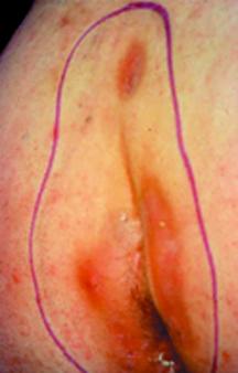
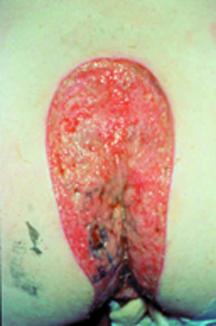
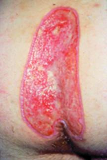
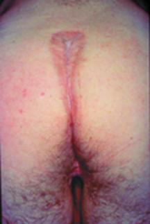
Wide excision and healing by secondary intention: Wide excision of an elliptical wedge of skin and subcutaneous tissue down to the pre-sacral fascia is designed to remove all the inflamed tissue and debris allowing the wound to granulate from its base. The excised dimensions should be of sufficient width at both the mouth and base of the wound to allow packing with ease. The base itself should be relatively flat and of almost comparable size to the mouth of the wound. A narrow V-shaped wound without a flat base is more difficult to pack and has a tendency to bridging and subsequent infection. The procedure necessitates general anaesthesia and hospital stay for a few days postoperatively. The principal advantage is a low recurrence rate but the downside is a lengthy healing time (8-10 weeks)[16] and high direct and indirect costs associated with inpatient care, follow-up wound care and days lost from work. Despite this there is a role for wide excision in those with extensive chronic disease and following failed primary closure surgical technique. Figures 1a-e show different stages in the healing of chronic disease in a young male treated by wide excision.
A modification of the standard excision is 'marsupialisation'. The skin edges are not excised, but are sutured to the sides of the wound. The mean healing time in 125 patients who underwent this procedure was shown to be four weeks with a recurrence rate of 6%[12].
Excision and primary closure: Closure of the wound is more cosmetically acceptable for some patients and is associated with a shorter healing time and time off work. However, this potential benefit is offset by the need for bed rest for up to one week in hospital[17] coupled with a higher risk of postoperative infection. When infection intervenes the wound must be laid open and healing time is longer than if the wound had been treated by secondary intention in the first place. The scar can be sited over the midline or displaced laterally with one year recurrence rates of 18% and 10% respectively[5]. In a recent prospective study failure of primary healing was significantly associated with early recurrence of disease[18]. In the same study the use of preoperative antibiotics did not influence the recurrence rate.
Bascom has proposed a method to incise, drain and curette a chronic abscess through a lateral incision combined with excision of any midline pits with a minimal amount of surrounding tissue[7]. A section of the wall of the abscess cavity opposite the incision is raised as a flap and used to close the communication between the midline pits and the abscess cavity. This is accomplished by suturing the flap to the underside of the skin bridge formed between the incision and the midline. In a recent study of 218 patients treated with Bascom's procedure as day cases, 6% developed a postoperative abscess requiring drainage and 10% had recurrence requiring further surgery at mean follow up of 12.1 months (range 1-60 months)[19].
Excision with reconstructive procedures: These procedures are more technically demanding and are probably best performed by a plastic surgeon. Their use is generally restricted to recurrent complex pilonidal disease. The theory behind the majority of procedures is to reshape and flatten the natal cleft to reduce friction, local warmth, moisture and hair accumulation. Karydakis pioneered a procedure raising a flap to overlap the midline with the scar sited to one side to reduce postoperative hair entry[20]. Alternative techniques use a flap of both skin and muscle or a Z-plasty flap to close the defect following excision[17]. All these techniques require general anaesthesia and a week or more of bed rest in hospital.
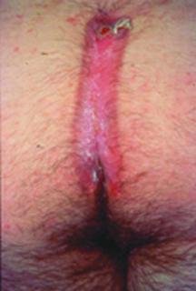
Following incision and drainage or excision procedures the wound should be packed with an alginate dressing. Following this the wound can be managed with foam dressing that forms in situ (eg Cavicare) and an appropriate secondary dressing. The foam dressing keeps the wound open preventing premature closure of the wound edges. However, a new stent needs to be made approximately every two weeks to allow for wound contracture and is contraindicated when there are signs of infection. Hair and any debris should be removed at every wound inspection and the natal cleft should be kept hair free by weekly shaving or use of depilatory agents. Figure 2 shows a postoperative wound with signs of bridging and insufficient attention to depilation. Depilation is usually continued until wound healing, although the authors would recommend this practice continue long term[21] or at least until the patient reaches his late thirties when recurrence is unlikely.
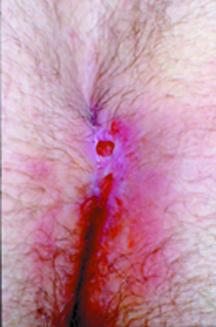
Early signs of wound infection include increased pain and an abnormal dark 'beefy' red appearance to the granulation tissue which is friable, bleeds on contact and exhibits superficial bridging. Prompt treatment with broad spectrum antibiotics such as metronidazole and erythromycin or clarithromycin is advised for a minimum of two weeks[10]. The use of dressings containing antiseptics such as iodine (eg Inadine), although controversial, may have beneficial effects.
Recurrence can be divided into two groups: early and late. Early recurrence is usually due to failure to identify one or more sinuses at incision and drainage, which was not followed by a second-look procedure. Late recurrence is usually due to secondary infection caused by residual hair or debris that was not removed at operation, inadequate wound care or insufficient attention to depilation[21].
Pilonidal disease is a complex condition that causes both discomfort and embarrassment to sufferers. Direct costs to the healthcare system and indirect costs through absence from work are high. Incision and drainage (with curettage) is recommended for treatment of pilonidal abscesses. Wide excision and healing by secondary intention is the recommended treatment of most chronic disease. The surgical management of complex or recurrent disease should be under a surgeon with an interest in pilonidal disease. Regardless of the surgical technique concerned, standard principles of wound care are essential with repeated depilation of the natal cleft, removal of hair and any debris from the wound bed and keeping the wound edges separated using an appropriate dressing.
1. Hodges RM. Pilonidal sinus. Boston Med Surg J 1880; 103: 485-586.
2. Sondenaa K, Nesvik I, Anderson E, Natas O, Soreide JA. Patient characteristics and symptoms in chronic pilonidal sinus disease. Int J Colorectal Dis 1995; 10(1): 39-42.
3. Dwight RW, Maloy JK. Pilonidal sinus: experience with 449 cases. N Engl J Med 1953; 249: 926-30.
4. Buie LA, Curtis PD. Pilonidal disease. Surg Clin NA 1952; 32: 1247-59.
5. Berry DP. Pilonidal sinus disease. J Wound Care 1992; 1(3): 29-32.
6. Price ML, Griffiths WAD. Normal body hair: a review. Clin Exp Dermatol 1985; 10: 87-97.
7. Bascom JU. Pilonidal disease: correcting over treatment and under treatment. Contemporary Surg 1981; 18: 13-28.
8. Lineaweaver WC, Brunson MB, Smith JF, Franzini DA, Rumley TO. Squamous carcinoma arising in a pilonidal sinus. J Surg Oncol 1984; 27(4): 39-42.
9. Trent JT, Kirsner RS. Wounds and malignancy. Adv Skin Wound Care 2003; 16(1): 31-34.
10. Marks J, Harding KG, Hughes LE, Riberio CD. Pilonidal sinus excision: healing by open granulation. Br J Surg 1985; 72(8): 637-40.
11. Sondenaa K, Nesvik I, Andersen E, et al. Bacteriology and complications of chronic pilonidal sinus treatment with excision and primary suture. Int J Colorectal Dis 1995; 10(3): 161-66.
12. Solla JA, Rothenberger DA. Chronic pilonidal disease. An assessment of 150 cases. Dis Col Rec 1990; 33(9): 758-61.
13. Hodgkin W. Pilonidal sinus disease. J Wound Care 1998; 7(9): 481-83.
14. Stansby G, Greatorex R. Phenol treatment of pilonidal sinus of the natal cleft. Br J Surg 1989; 76(7): 729-30.
15. Bissett IP, Isbister WH. The management of patients with pilonidal disease: a comparative study. Aust NZ J Surg 1987; 57(12): 939-42.
16. Kronberg I, Christensen KI, Zimmerman-Nielson O. Chronic pilonidal disease: a randomised trial with complete three year follow up. Br J Surg 1986; 72: 303-04.
17. Jones DJ. ABC of colorectal diseases. Pilonidal sinus. BMJ 1992; 305: 410-12.
18. Sondenaa K, Diab R, Nesvik I, et al. Influence of failure of primary wound healing on subsequent recurrence of pilonidal sinus. Eur J Surg 2002; 168(11): 614-18.
19. Senapati A, Cripps NP, Thompson MR, Franzini DA. Bascom's operation in the day-surgical management of symptomatic pilonidal sinus. Br J Surg 2000; 87: 1067-70.
20. Karydakis GE. New approach to the problem of pilonidal sinus. Lancet 1973; 2: 1414-15.
21. Allen-Marsh TG. Pilonidal sinus: finding the right track for treatment. Br J Surg 1990; 77: 123-32.